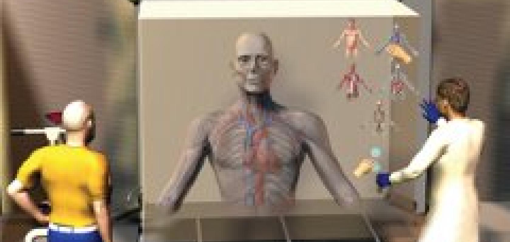
Researchers at the Institute for Biomedical Imaging and Modelling (INSIGNEO), a new research institute set up by the University of Sheffield and Sheffield Teaching Hospitals NHS Foundation Trust, are working towards building a patient-specific avatar based on an individual’s medical data. The researchers are currently developing computer models of different organs, including models of the cardiac and neuro-musculo-skeletal systems, but the ultimate aim is to build a complete digital replica of a patient.
The computer models will incorporate medical information such as age and weight, as well as X-ray and other medical imaging data to provide an overall picture of the patient’s condition, against which different treatments can be tested.
The benefit is better targeted treatments for the patient. Certain treatments might have a low success rate, so being able to simulate their likely outcome on a patient avatar will help clinicians decide on treatment options.
In a statement released at a press conference to launch UK National Science and Engineering week, director of INSIGNEO, Professor Alejandro Frangi, said: ‘By developing models of complete organ systems, such as the cardiovascular system, we can help clinicians predict whether treating a constriction in one coronary artery, for example, might improve or worsen blood supply in other coronary arteries in patients with multiple lesions. Because it’s currently impossible to make these kinds of predictions, clinicians are often forced to deal with one issue at a time in diseases that are in fact multifactorial or systemic. Our models will enable doctors to handle illness in a more holistic way.’
Professor Frangi and his team are currently working on a model of a cerebral aneurysm, already being piloted with patients, that supports clinicians in predicting the likelihood of rupture, when treatment is necessary and what sort of treatment will work best. The model is based on data from X-ray or MRI scans showing the shape of the aneurysm and blood flow dynamics.
Other examples include a model of the heart’s aortic valve to help clinicians decide, in the case of heart valve failure, when a valve will need repair without the need for invasive tests. Also, a musculo-skeletal model to help predict the likelihood of bone fracture in elderly patients based on bone density data from scans and gait analysis.
Dr Julian Gunn, a cardiologist and senior lecturer at the department of cardiovascular science at the University of Sheffield, explained at the press conference that a simulation tool would allow cardiologists to decide on where best to position a stent in an artery and whether it will extend the patient’s life. Dr Gunn stated about the potential for the technology: ‘It will take us from the 19th to 22nd century in one go.’
Professor Frangi said much of the simulations are based on software tools used in the aerospace industry, such as that from Ansys. The critical issue, he said, is how to personalise those computer models.
In terms of modelling stents, Dr Gunn said this technology will be available soon, but Professor Frangi recognised that generating an entire virtual body is still a way off in the future, giving a timeframe of 20 years or more.