Three cardiac anaesthetists practicing at the Heart Hospital in London (University College London Hospitals) have teamed up with digital animation specialists, Glassworks, to produce a beating 3D computer-generated model of the human heart. The brainchild of Dr Sue Wright, Dr Andrew Smith, and Dr Bruce Martin, all of UCLH Heart Hospital, the HeartWorks project was developed as a teaching tool for peri-operative transoesophageal echocardiography (TOE), or cardiac ultrasound.
The model uses Nvidia's Quadro GPU (graphics processing unit) to provide the necessary processing power to simulate a real-time beating heart using a relatively low power CPU. California-based Nvidia is a provider of visual computing technologies for the entertainment, design, and high-performance computing markets. Adam Cubitt of Glassworks explained that Nvidia's graphics card allowed the simulation to render images quickly enough to run at the 30fps required to make the animation realistic, avoiding it skipping as it moved from one frame to the next.
Prior to heart surgery, the TOE procedure is carried out by passing an ultrasound probe down the patient's throat and into the stomach, to rest just below the patient’s heart. The ultrasound pictures provide the anaesthetist with vital clues about the condition of the cardiac muscles.
Training aids of the heart are generally based on textbook diagrams, such as those found in Gray's Anatomy, or on plastic models, and learning where to place the probe during a TOE is difficult to teach. Most anaesthetists learn the procedure through on-the-job training. 'It can take weeks or months to associate the 2D structure on the ultrasound screen with a 3D image of the heart,' commented Wright of UCLH Heart Hospital.
The goals of the HeartWorks project were to create an interactive teaching tool that was anatomically accurate, that was animated to beat in real-time to show changes in the heart's shape during the cardiac cycle, and that incorporated an ultrasound tool.
The team at Glassworks created the animation by collating huge amounts of digital images of the heart. Cubitt of Glassworks noted that working with the clinicians at UCLH Heart Hospital provided the Glassworks team with access to the best datasets and to comment from top experts in the field.
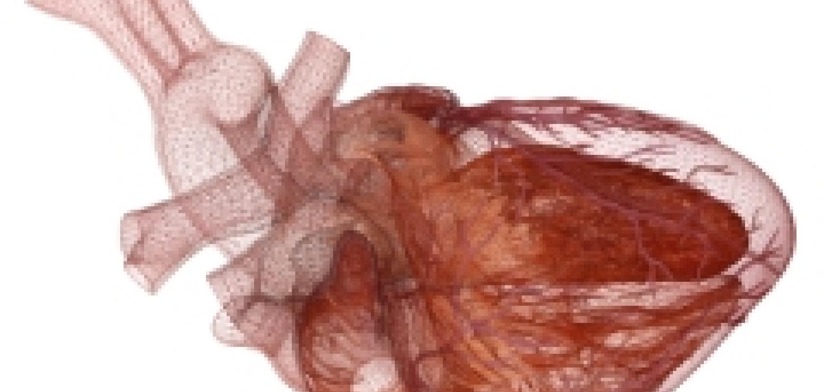
The starting point was an optical scan of an interior cast of a healthy heart, which was supplemented with masses of other reference material. The model is a top-down construction of a heart, rather than building it up from a cellular level and animating the different cells and tissues working together. Cubitt explained that the aim was to create a simulation that's not a perfect model of the heart, but such a good replica that everyone will believe it is.
The model incorporates ultrasound simulation, with a probe and mannequin providing a hands-on teaching tool for the TOE procedure. Trainee anaesthetists can compare the ultrasound images of the heart to the plane of the ultrasound slicing through the animated 3D model.
The model can be rotated and viewed from different angles, as well as zooming in on different structures. 'There is the ability to remove certain features that block the view of what's being studied,' explained Wright of UCLH Heart Hospital. 'Surgeons get a view of the heart they'd never normally see,' she said, such as the valves opening and closing. A textbook-based feature is also available, where users can click on an anatomical structure in the model to bring up a textbook description of the structure in question. The GPU from Nvidia allowed the Glassworks team to create such a dynamic and interactive model.
The HeartWorks project was funded by the University College London Hospitals Charity and was launched in May 2008. The mannequin simulator is commercially available from September 2008.
